一套荧光检漏套装解决工业设备泄漏
LUYOR-6801荧光检漏仪是美国路阳公司生产的荧光检漏仪套装,适用于检查小到中等规模的油基液体系...
2024-03-28

作者:时间:2019-09-05 10:26浏览12157 次
mCherry是一种来自于蘑菇珊瑚(mushroom coral)的红色荧光蛋白,常有于标记和示踪某些分子和细胞组分。相对于其他荧光,mCherry的好处在于它的颜色和应用最多的绿色荧光蛋白(GFP)能进行共同标记,并且mCherry相对于其他单体荧光蛋白来说也具有卓越的光稳定型。
mcherry激发波长是多少?
mCherry是一种来自于蘑菇珊瑚(mushroom coral)的红色荧光蛋白,常有于标记和示踪某些分子和细胞组分。相对于其他荧光,mcherry的好处在于它的颜色和应用最多的绿色荧光蛋白(GFP)能进行共同标记,并且mcherry相对于其他单体荧光蛋白来说也具有卓越的光稳定型。

吸收和发射波长:
mcherry的更大吸收/发射峰分别位于587nm和610nm,对光致漂白耐受,荧光非常稳定。

常见应用:
•mCherry常用于与目的基因组成融合蛋白以及通过IRES或2A与感兴趣的蛋白共表达;
•启动子活性研究;
•荧光共振能量转移(fluorescence resonance energy transfer,FRET)和其他定量实验;
•标记细胞或者分子,进行示踪实验;

mCherry is a fluorophore (a fluorescent protein) used in biotechnology as a tracer to follow the flow of fluids, as a marker when tagged to molecules and cell components. mCherry and the majority of red fluorescent proteins derive from a protein isolated from Discosoma sp., while other fluorescent proteins in the green range are often variants of GFP from Aequorea victoria.
m前缀的意义:
mCherry的m为monmer单体的缩写,表示mCherry荧光蛋白的形式为单体,这在很多实验设计中非常重要,比如与目的标记基因组成融合蛋白时。
成熟时间:
mCherry具有较快的成熟速度,t0.5为15分钟,这在一些需要做出快速反应的实验非常重要。比如启动子活性报告系统等。
如何观察和筛选mcherry在植物中的表达?
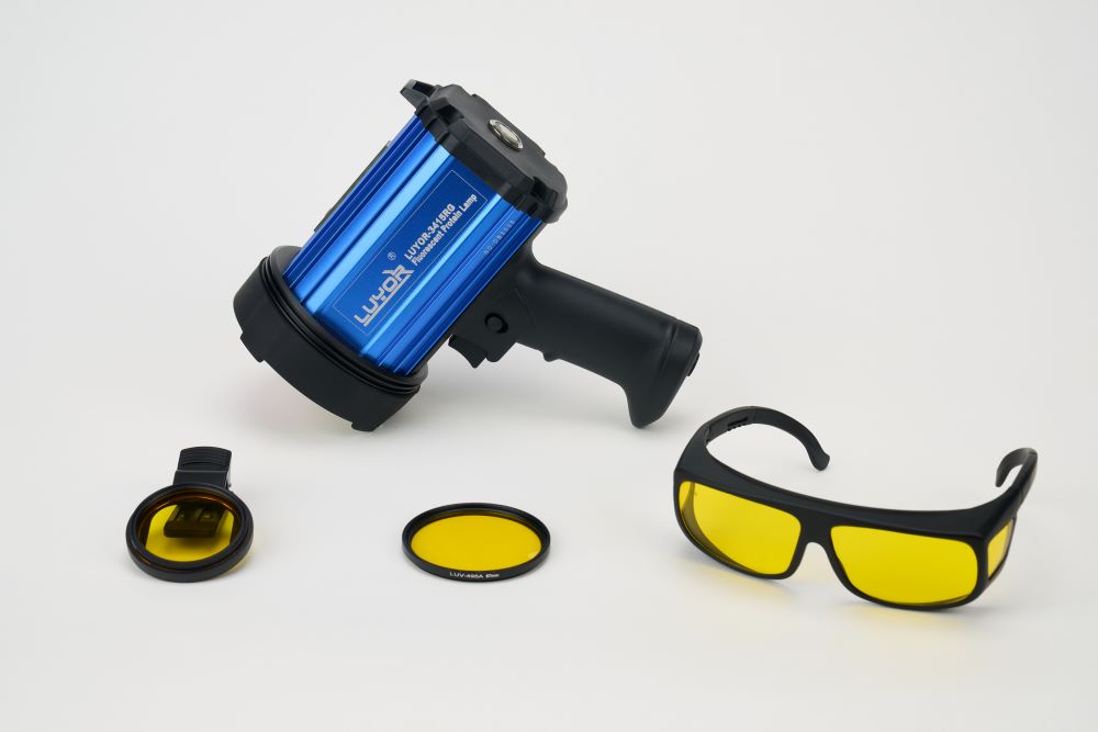
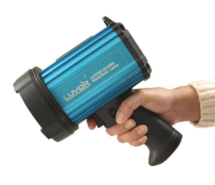
你可以选用LUYOR-3415RG和LUYOR-3260GR便携式荧光蛋白激发光源来直接观察和筛选mcherry在植物中有没有表达。如需进一步了解,请拨打电话153-1756-5658进行咨询(或直接添加微信15317565658咨询)。

mcherry红色荧光蛋白的激发波长和发射波长

上图为LUYOR-3415RG便携式红色荧光蛋白激发光源

上图为LUV-50A红色荧光蛋白观察眼镜

上图为LUV-590A 红色荧光蛋白拍照滤镜

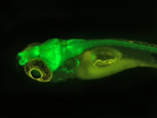
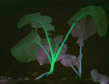
上图为红色荧光蛋白在大豆根系上的表达(LUYOR-3415RG照射,LUV-590A滤镜拍摄)
实验室常见荧光蛋白的激发波长和发射波长
紫外荧光蛋白UV Proteins
| Protein | Excitation Wavelength | Emission Wavelength |
|---|---|---|
| Sirius | 355 | 424 |
| Sandercyanin | 375 | 630 |
| shBFP-N158S/L173I | 375 | 458 |
蓝色荧光蛋白Blue Proteins
| Protein | Excitation Wavelength | Emission Wavelength |
|---|---|---|
| Azurite | 383 | 450 |
| EBFP2 | 383 | 448 |
| mKalama1 | 385 | 456 |
| mTagBFP2 | 399 | 454 |
| TagBFP | 402 | 457 |
| shBFP | 401 | 458 |
青色荧光蛋白Cyan Proteins
| Protein | Excitation Wavelength | Emission Wavelength |
|---|---|---|
| ECFP | 433 | 475 |
| Cerulean | 433 | 475 |
| mCerulean3 | 433 | 475 |
| SCFP3A | 433 | 474 |
| CyPet | 435 | 477 |
| mTurquoise | 434 | 474 |
| mTurquoise2 | 434 | 474 |
| TagCFP | 458 | 480 |
| mTFP1 | 462 | 492 |
| monomeric Midoriishi-Cyan | 470 | 496 |
| Aquamarine | 430 | 474 |
绿色荧光蛋白Green Proteins
| Protein | Excitation Wavelength | Emission Wavelength |
|---|---|---|
| TurboGFP | 482 | 502 |
| TagGFP2 | 483 | 506 |
| mUKG | 483 | 499 |
| Superfolder GFP | 485 | 510 |
| Emerald | 487 | 509 |
| EGFP | 488 | 507 |
| Monomeric Azami Green | 492 | 505 |
| mWasabi | 493 | 509 |
| Clover | 505 | 515 |
| mNeonGreen | 506 | 517 |
| NowGFP | 494 | 502 |
| mClover3 | 506 | 518 |
黄色荧光蛋白Yellow Proteins
| Protein | Excitation Wavelength | Emission Wavelength |
|---|---|---|
| TagYFP | 508 | 524 |
| EYFP | 513 | 527 |
| Topaz | 514 | 527 |
| Venus | 515 | 528 |
| SYFP2 | 515 | 527 |
| Citrine | 516 | 529 |
| Ypet | 517 | 530 |
| lanRFP-ΔS83 | 521 | 592 |
| mPapaya1 | 530 | 541 |
| mCyRFP1 | 528 | 594 |
桔色荧光蛋白Orange Proteins
| Protein | Excitation Wavelength | Emission Wavelength |
|---|---|---|
| Monomeric Kusabira-Orange | 548 | 559 |
| mOrange | 548 | 562 |
| mOrange2 | 549 | 565 |
| mKOκ | 551 | 563 |
| mKO2 | 551 | 565 |
红色荧光蛋白Red Proteins
| Protein | Excitation Wavelength | Emission Wavelength |
|---|---|---|
| TagRFP | 555 | 584 |
| TagRFP-T | 555 | 584 |
| RRvT | 556 | 583 |
| mRuby | 558 | 605 |
| mRuby2 | 559 | 600 |
| mTangerine | 568 | 585 |
| mApple | 568 | 592 |
| mStrawberry | 574 | 596 |
| FusionRed | 580 | 608 |
| mCherry | 587 | 610 |
| mNectarine | 558 | 578 |
| mRuby3 | 558 | 592 |
| mScarlet | 569 | 594 |
| mScarlet-I | 569 | 593 |
远红荧光蛋白Far Red Proteins
| Protein | Excitation Wavelength | Emission Wavelength |
|---|---|---|
| mKate2 | 588 | 633 |
| HcRed-Tandem | 590 | 637 |
| mPlum | 590 | 649 |
| mRaspberry | 598 | 625 |
| mNeptune | 600 | 650 |
| NirFP | 605 | 670 |
| TagRFP657 | 611 | 657 |
| TagRFP675 | 598 | 675 |
| mCardinal | 604 | 659 |
| mStable | 597 | 633 |
| mMaroon1 | 609 | 657 |
| mGarnet2 | 598 | 671 |
近红荧光蛋白Near IR Proteins
| Protein | Excitation Wavelength | Emission Wavelength |
|---|---|---|
| iFP1.4 | 684 | 708 |
| iRFP713 (iRFP) | 690 | 713 |
| iRFP670 | 643 | 670 |
| iRFP682 | 663 | 682 |
| iRFP702 | 673 | 702 |
| iRFP720 | 702 | 720 |
| iFP2.0 | 690 | 711 |
| mIFP | 683 | 704 |
| TDsmURFP | 642 | 670 |
| miRFP670 | 642 | 670 |
Sapphire-type Proteins
| Protein | Excitation Wavelength | Emission Wavelength |
|---|---|---|
| Sapphire | 399 | 511 |
| T-Sapphire | 399 | 511 |
| mAmetrine | 406 |
526 |
Red fluorescent proteins (RFP) can be imaged on existing confocal or widefield microscopes, and they also have more penetrating power. The excitation and emission maxima of RFP are 558nm and 583 nm, respectively.
The use of RFP, however, has been hampered with several issues. RFP is an obligate tetramer - thus, it forms large aggregates inside cells. This makes the use to RFP to report the location of a protein severely limited.
Although GFP can successfully fuse with several hundreds of proteins, RFP-conjugated proteins are often toxic. Some variants of RFP have overcome these limitations. For example, DsRed2 fluorescent protein does not form aggregates and has reduced toxicity, while another variant of RFP (known as RedStar) has increased brightness and maturation rate.
 关注我们
关注我们